Surgical stitches, or sutures, are used to close wounds following injury or surgery and to support the healing process. But sutured wounds are susceptible to infection, with infections at surgical sites occurring in 2–4% of patients.
A team headed up at RMIT University in Australia is developing an antimicrobial suture that could play an important role in preventing such infections. The smart suture is also designed to be visible in CT scans, enabling wound monitoring after surgery and enhancing patient recovery. This could prove particularly useful for locating internal stitches and to help detect infected wounds inside the body without requiring surgical intervention.
“Our smart surgical sutures can play an important role in preventing infection and monitoring patient recovery,” says lead author Shadi Houshyar in a press statement. “The proof-of-concept material we’ve developed has several important properties that make it an exciting candidate for this.”
Currently, there are no commercial contrast agents that can be used in sutures due to problems with toxicity. To get around this, Houshyar and colleagues are using iodine-conjugated carbon particles (ICPs). Carbon nanoparticles are biocompatible, cheap and easy to produce in the lab, and inherently fluorescent and antibacterial. Attaching iodine provides X-ray visibility and enhanced antimicrobial properties, while lowering the iodine toxicity by controlling the release of ICPs. The team embedded these ICPs into the suture material polycaprolactone (PCL) to create I-PCL suture filaments.
Describing their work in OpenNano, the researchers first evaluated the stability of the I-PCL before complete suture degradation. They immersed sutures in phosphate-buffered saline (PBS) at 37 °C for 22 days with mild shaking and quantified the iodine concentration in the PBS. In the first 24 hrs, they saw an initial high-rate release of ICP. This was followed by a significant drop in ICP concentration and slow, steady release over time.
This burst release of ICPs from the suture could prevent the colonization of wounds with pathogens, while the ensuing slow release over an extended time period helps prevent biofilm formation and infection.
The team then examined the impact of ICP concentration on the contrast properties of I-PCL, comparing micro-CT images of sutures containing various ICP concentrations. The CT contrast increased with increasing ICP concentration. The suture with 40% ICP (40I-PCL) exhibited contrast 272, 81 and 31% higher than sutures with 10, 20 and 30% ICP concentrations, respectively.
Focusing on the 40I-PCL suture with the highest contrast enhancement, the researchers investigated how suture degradation affected its contrast properties. After 22 days in PBS, the CT contrast reduced by 18%, which the team notes is still acceptable and as expected for a degradable suture.
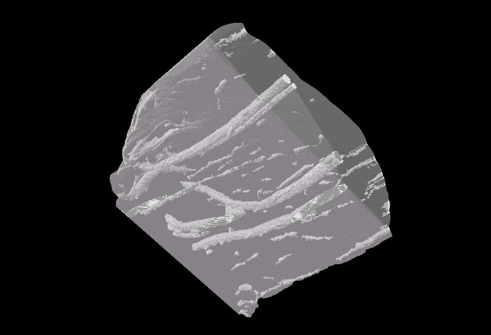
The researchers next assessed the visibility of 40I-PCL sutures threaded through chicken breast and thigh tissue. Micro-CT images revealed that sutures were clearly visible in, and distinguishable from, the tissue. Such visibility should be particularly advantageous for post-surgery wound monitoring, enabling surgeons to identify the suture integrity and location within the body.
Antibacterial optimization
The bacteria methicillin-resistant S. aureus (MRSA) is the most common cause of surgical site infections, thus the researchers studied its interaction with various I-PCL sutures. After 6 h incubation at 37°C, 30I-PCL killed 90% of MRSA, while the higher-concentration 40I-PCL eliminated nearly 99% of MRSA with no visible biofilm on the suture.
In addition to use in surgical stitches, the smart suture material can also be employed to create meshes, such as the vaginal mesh implants used to treat prolapse – a procedure that can have relatively high infection rates.

Photosynthetic sutures promote wound healing
“This mesh will enable us to help with improved identification of the causes of symptoms, reduce the incidence of mesh infections and will help with precise pre-operative planning if there is a need to surgically remove the mesh,” explains co-author Justin Yeung from the University of Melbourne. “It has the potential to improve surgery outcomes and improve quality-of-life for a huge proportion of women, if used as vaginal mesh for example, by reducing the need for infected mesh removal.”
The researchers conclude that sutures that can be visualized using modalities such as CT and MRI can minimize surgical risks, monitor internal wounds and assist surgeons accurately planning surgery if complications requiring mesh removal develop. They suggest that the 40I-PCL suture best satisfies these requirements by providing high CT visibility, as well as reasonable biocompatibility and excellent antimicrobial properties.
- SEO Powered Content & PR Distribution. Get Amplified Today.
- Platoblockchain. Web3 Metaverse Intelligence. Knowledge Amplified. Access Here.
- Source: https://physicsworld.com/a/smart-stiches-could-prevent-infections-at-surgical-sites/