Spaceflight – both short-duration space shuttle missions and longer periods living on the International Space Station (ISS) – alters the human body, including widespread changes in the brain. By studying brain scans of astronauts before and after space travel, a multi-institutional research team has found that ventricles – fluid-filled cavities in the brain – expand significantly in longer spaceflight missions, and that inter-mission intervals of less than three years may not be long enough for them to fully recover.
Time spent in space induces displacement of intracranial fluid and an upward shift of the brain within the skull, causing cortical crowding and narrowing of the sulci, the grooves in the cerebral cortex, at the top of the brain. With future space exploration including more long-duration missions, it is imperative to understand the effects of space travel on the brain.
With this goal, Rachael Seidler and Heather McGregor of the University of Florida and co-researchers studied the brains of 30 astronauts, evaluating changes in grey matter (GM) volume, white matter (WM) microstructure, extra-cellular free water (FW) distribution and ventricular volume from before to after spaceflights. The group included both novices and experienced astronauts, on missions ranging from two weeks to 12 months.
In a prospective study conducted between 2014 and 2020, the researchers obtained pre- and post-flight data (T1-weighted and diffusion-weighted MRI scans) from 13 astronauts who spent six months and two who spent 12 months in space. They also examined retrospective MRI data from the NASA Lifetime Surveillance of Astronaut Health Repository. This included eight astronauts who completed a short-duration mission of up to two weeks, and seven who spent six to 12 months on the ISS.
The goal of the study was to understand whether longer missions or shorter recovery periods between missions have a particularly significant impact on the brain. The team examined whether and how spaceflight-induced brain changes are associated with individual differences in spaceflight experience, factoring in the duration of the mission, the experience level of the astronaut, the number of previous missions and the time elapsed since a previous mission.
The researchers compared pre- and post-flight diffusion-weighted MRI data for the astronauts, assessing group-level changes in GM volume, ventricular volume, FW fractional volume, and FW-corrected WM diffusion indices. They developed models that adjusted for individual differences in age at the time of launch, sex, current mission duration, and the number of days between landing and the post-flight MRI scan. For each astronaut, they calculated FW difference images reflecting changes in FW fractional volume and performed ventricular volume analysis.
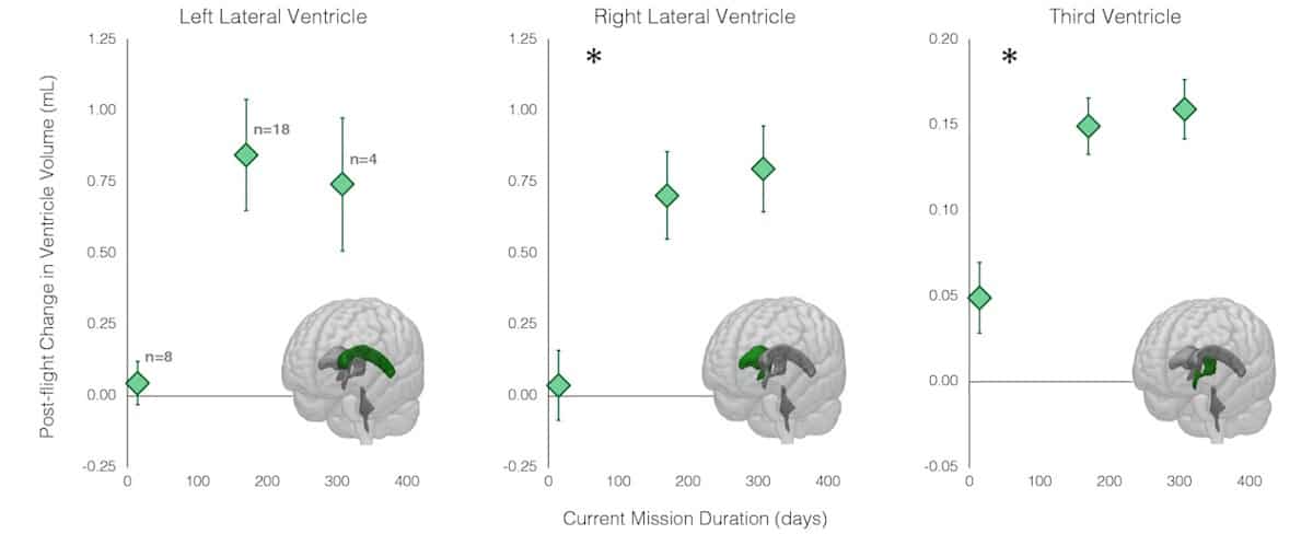
These analyses demonstrated that ventricular volume increased during spaceflight, with longer missions resulting in greater ventricular enlargement that tapered off after six months in space. The study did not resolve the length of time for the ventricles to fully recover post-flight, or to what extent they recover. But based on the findings, described in Scientific Reports, the team suggests that at least three years or longer intervals between spaceflight missions are needed for intracranial fluid to return to normal levels and for the ventricles to fully recover.
The study also confirmed previous published findings that spaceflight induces grey matter shifts and FW redistribution. The researchers determined that longer missions induce greater fluid shifts. Among the experienced astronauts, the number of years elapsed since the previous mission was significantly associated with post-flight volume changes for all four ventricles.
Seidler and colleagues point out that the rate of ventricular expansion during spaceflight exceeds that seen with normal aging, suggesting that post-flight ventricular enlargement is not a consequence of brain atrophy with aging, but instead arises from cerebrospinal fluid changes. They suggest that this enlargement may be a compensatory response to accommodate fluid shifts toward the head that occur in microgravity.
Blood tests reveal brain damage following long-term spaceflight
The researchers note that variation in astronaut age did not account for observed individual differences in structural brain changes, and that there were no differences in spaceflight-induced structural brain changes between novice and experienced astronauts. Rather, brain changes differed according to the number of prior flights the experienced astronauts had completed. “This suggests that the brain is impacted by the cumulative effects across multiple flights and perhaps separate bouts of adaptation to microgravity and the spaceflight environment,” they write.
“Collectively, our work suggests that longer missions, multiple flights and shorter inter-mission recovery time induce greater intracranial fluid changes. Moreover, the findings suggest that neuroplasticity changes resulting from adaptation to microgravity may not be dependent on previous spaceflight experience,” they conclude. Seidler tells Physics World that the team is now conducting a new study with long-term post-flight follow up, up to five years after a mission.
- SEO Powered Content & PR Distribution. Get Amplified Today.
- PlatoData.Network Vertical Generative Ai. Empower Yourself. Access Here.
- PlatoAiStream. Web3 Intelligence. Knowledge Amplified. Access Here.
- PlatoESG. Automotive / EVs, Carbon, CleanTech, Energy, Environment, Solar, Waste Management. Access Here.
- BlockOffsets. Modernizing Environmental Offset Ownership. Access Here.
- Source: https://physicsworld.com/a/life-in-space-impacts-human-brain-structure/